
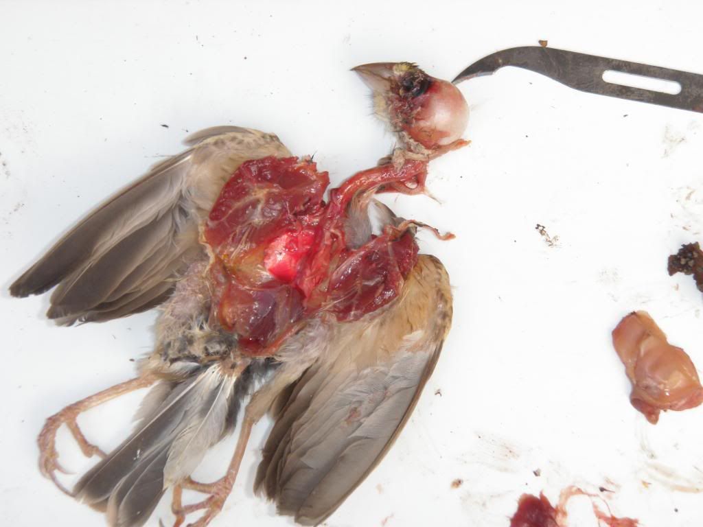
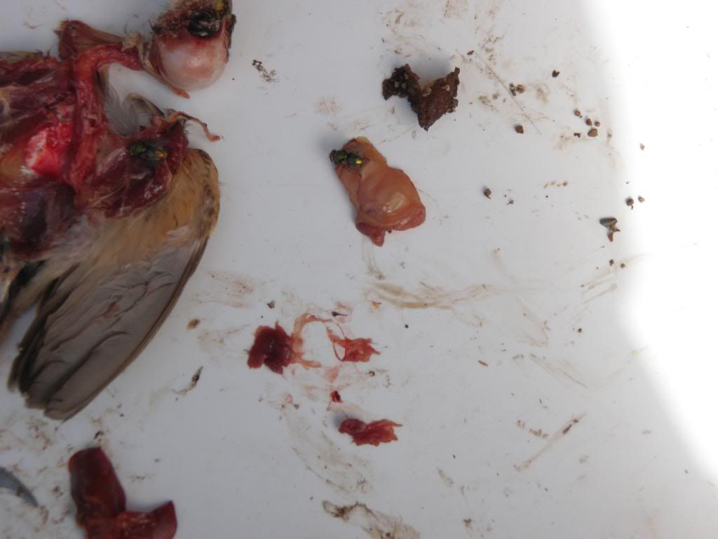
Last week, on a very hot day, I noticed my hen weaver was in a distressed state. The following day she was observed in a similar condition. I grabbed my catching net and a holding box, unfortunately by the time I located the hen in the planted aviary she had died. A visual check of the corpse revealed the weaver was a 4 to 5 year old hen that was emaciated,with signs of diarrhoea and a small haematoma on the skull.
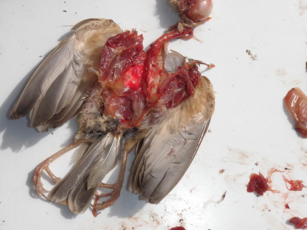
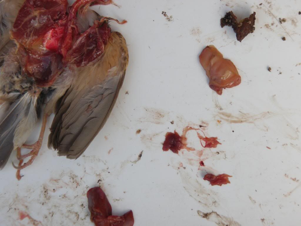
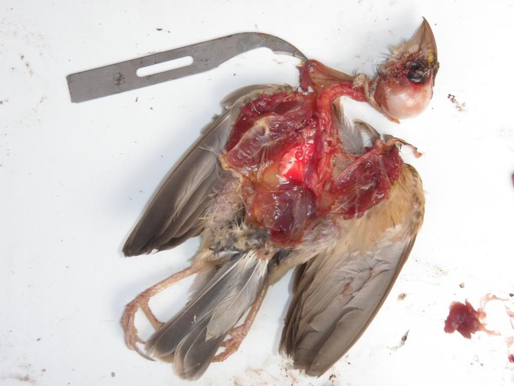
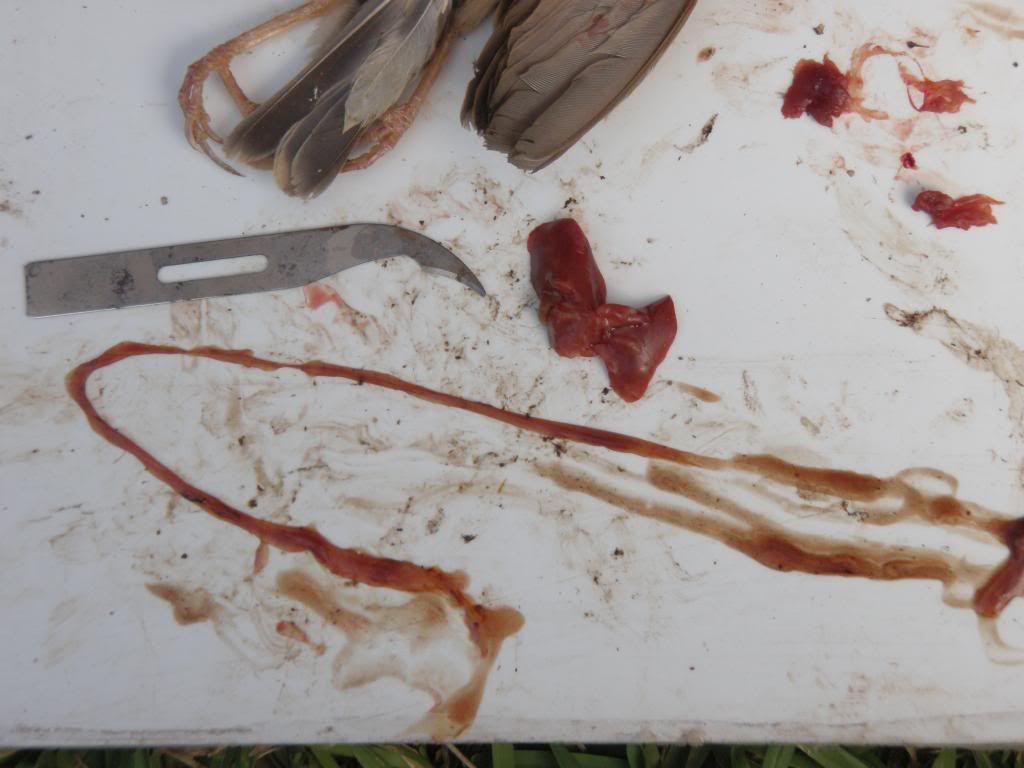
The following photos are of the hen weaver after I had completed the autopsy. Only instruments used was a stitch cutter and a pair of scissors.
http://i925.photobucket.com/albums/ad93 ... 09af85.jpg
http://i925.photobucket.com/albums/ad93 ... b8b844.jpg
http://i925.photobucket.com/albums/ad93 ... 0e4a07.jpg
stitch cutter points towards are of damage found on skull
http://i925.photobucket.com/albums/ad93 ... a1fc2c.jpg
http://i925.photobucket.com/albums/ad93 ... 09bf2b.jpg
http://i925.photobucket.com/albums/ad93 ... f69edb.jpg
my assistants during the 12 minute procedure - the fly's
http://i925.photobucket.com/albums/ad93 ... f97ca6.jpg
Any thoughts?
Found this hen weave dead in aviary -Autopsy photos attached
-
mattymeischke

- ...............................

- Posts: 862
- Joined: 25 Jul 2011, 20:25
- Location: Southern Tablelands of NSW
Hi, Wagga, sorry 'bout your hen.
I can't glean much from the photos; it is very different looking at post-mortem photos compared with doing the dissection and it is hard to see which bits are where.
However, my two points would be:
(i) Small scalp haematomata are common post-mortem findings and often represent pooling of blood that has happened after the bird has already died, so it is hard to draw conclusions from that.
A large haematoma with an associated skull fracture would be good evidence for death by head injury.
(ii) If diarrhoea and emaciation were the most obvious clinical signs, then microscopic examination of faeces may be more helpful than gross examination; if a treatable pathogen were found this would also be important for the health of your surviving birds.
It is possible to take a faecal specimen post-mortem, though it is probably too late in this case. Otherwise, a pooled faecal specimen from your (living) flock could be sent for examination.
Usual caveats apply: I am not a vet, and have limited experience.
Those are my thoughts....
Avid amateur aviculturalist; I keep mostly australian and foreign finches.
The art is long, the life so short; the critical moment is fleeting and experience can be misleading, crisis is difficult....... (Hippocrates)
The art is long, the life so short; the critical moment is fleeting and experience can be misleading, crisis is difficult....... (Hippocrates)
-
wagga
- ...............................

- Posts: 678
- Joined: 24 Apr 2010, 22:08
- Location: Port Macquarie NSW 2444
- Location: PORT MACQUARIE NSW
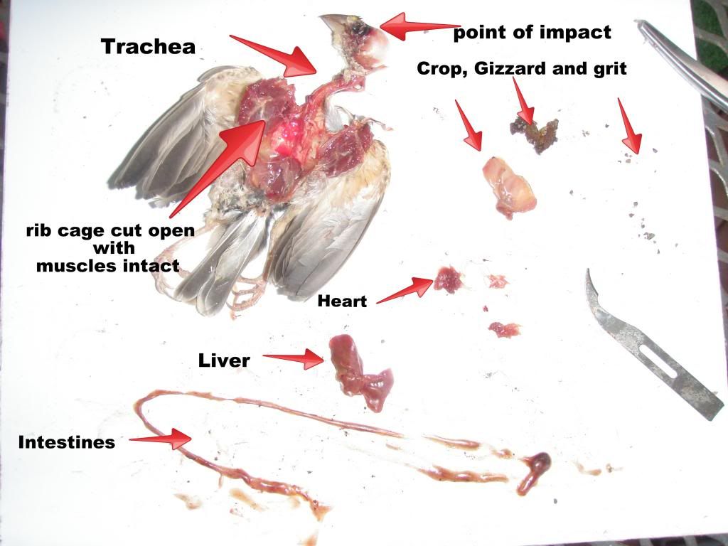
On reflection, Yes, I do agree that unless labelled correctly in a step by step sequence these photos are just poor quality photos of a dead bird. I will try and edit/label these photos which might help others recognise the bird's disemboweled organs on display.
Unfortunately I found the bird hours prior to travelling up to Armidale for the week and due to this time constraint, I was unable collect a faecal sample or send a tissue sample to a vet at the time.
[Usual caveats apply: I am not a vet, and have limited experience.
Those are my thoughts.] Thanks Matt for your valued opinion.
Al
Life in Port Macquarie is the ultimate Aussie sea change lifestyle.
-
wagga
- ...............................
- Posts: 678
- Joined: 24 Apr 2010, 22:08
- Location: Port Macquarie NSW 2444
- Location: PORT MACQUARIE NSW
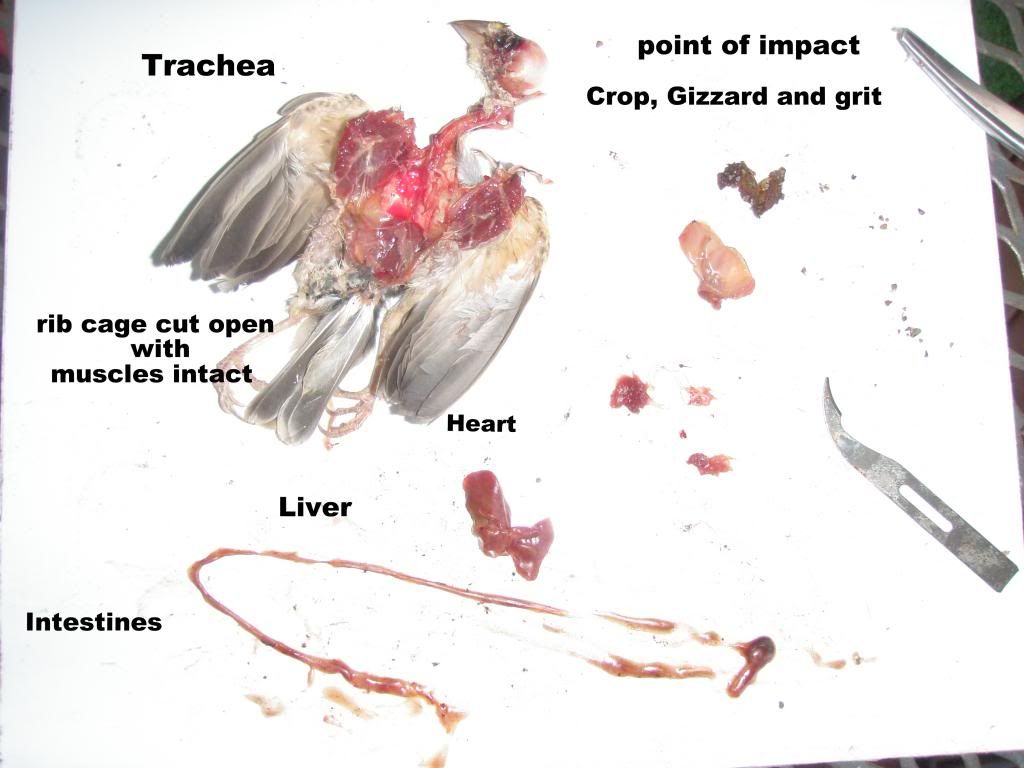
Here is a photo labelling the organs of the bird.
http://i925.photobucket.com/albums/ad93 ... 078d1d.jpg
BTW my wife will not look at this photo or even listen to my reasons why I have placed it this forum. I say "its for scientific and educational purposes". What do you think?
http://i925.photobucket.com/albums/ad93 ... 078d1d.jpg
BTW my wife will not look at this photo or even listen to my reasons why I have placed it this forum. I say "its for scientific and educational purposes". What do you think?
Life in Port Macquarie is the ultimate Aussie sea change lifestyle.

-
Myzomela

- ...............................

- Posts: 1545
- Joined: 24 Jan 2011, 18:44
- Location: Melbourne Vic
Sorry Wagga,
I can't really add any more to what Matty has already said; unfortunately it is really hard to see much because of the size of the organs but thanks for going to all the effort & labelling that last photo.
The only other thing that might help is to take photos of the organs inside the bird before they are dissected out ie take one with the belly skin and keel removed but the organs left in.
Taking macro shots may also reveal some more detail.
Hopefully you don't have a need to post any more photos any time soon, but weather extremes always find out the sick and elderly, nature's culling if you like.
Cheers
Myzo
Research; evaluate;observe;act
-
wagga
- ...............................
- Posts: 678
- Joined: 24 Apr 2010, 22:08
- Location: Port Macquarie NSW 2444
- Location: PORT MACQUARIE NSW
Thanks guys. I have learnt a very important lesson here. When in doubt, some things should be left to the professionals, especially if you want a answer why the bird died.
AL
AL
Life in Port Macquarie is the ultimate Aussie sea change lifestyle.
-
Diane
- ..............................

- Posts: 7402
- Joined: 05 Apr 2009, 14:23
- Location: Northern 'burbs of Adelaide
- Location: Northern 'burbs of Adelaide
While I can understand her reaction, its the only way I would ever get to see the inside of a bird. So I agree with your "scientific and educational purposes" statement.wagga wrote:BTW my wife will not look at this photo or even listen to my reasons why I have placed it this forum. I say "its for scientific and educational purposes". What do you think?
Diane
The difference between Genius and Stupidity is, Genius has it’s limits
The difference between Genius and Stupidity is, Genius has it’s limits